amfAR’s Dr. Rowena Johnston talks to Dr. Timothy Henrich about his use of advanced imaging technology to pinpoint hidden reservoirs of HIV.
Sun Tzu told his readers 2,500 years ago that in order to win a war, we must know our enemy. But the HIV reservoir—the collection of infected cells that persists even with antiretroviral therapy—has resisted efforts to reveal its essential nature. Understanding how much persists, and where, are fundamental challenges for researchers hoping to clear the reservoir, and ultimately to cure HIV.
While taking blood samples from people living with HIV is fairly easy, the reservoir in the blood is only a tiny fraction of the total, and is probably not representative of reservoir in tissues. Moreover, taking tissue samples is far more difficult and does not solve two crucial questions: Have the right pieces of tissue been taken? And does the reservoir behave the same way in a petri dish as it does in a person’s body?

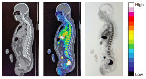
EXPLORER Imaging: (Left to right) CT scan, PET overlaid on CT, and PET image. Scale at right shows virus signal intensity.
To answer these questions, Dr. Tim Henrich and colleagues at the amfAR Institute for HIV Cure Research have turned to high-sensitivity PET imaging. They have forged a collaboration with researchers at the University of California, Davis to produce full-body images of people living with HIV, using the only EXPLORER scanner in the United States. The machine produces images that are approximately 40 times more sensitive than current technology, and in a fraction of the time.
Dr. Johnston is amfAR vice president and director of research.